 |
Jared Medina
Assistant Professor
Department of Psychology University of Delaware Email: jmedina AT psych DOT udel DOT edu |
|

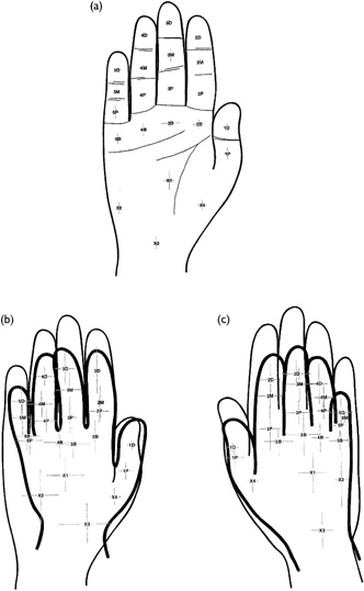
Although easily taken for granted, the coherent percept of our body can be altered due to impairment. Evidence from neurologically impaired individuals can inform us regarding how we integrate information from touch and other senses to create representations of the body. Previous studies with non-human primates observed cortical plasticity after brain damage, such that lesioned areas that previously represented a region of skin surface would reemerge in neighboring cortex - strong evidence for cortical plasticity and reorganization. However, as this research was limited to non-human primates, little systematic work had examined the sensory consequences of such changes. For example, after cortical reorganization, would someone localize touch correctly? Or would their sensation reflect some aspects of their pre-lesion experience? We examined an individual with extensive damage to the traditional "hand area" of primary somatosensory cortex. We found intact detection, suggesting cortical reorganization of the hand into other, intact areas of the brain. However, we also observed systematic errors in localization of tactile stimuli presented to the hand; perceived locations maintained the relative topography of the pre-lesion experience, but were shifted towards the center of the hand and away from the fingers (Rapp, Hendel, & Medina, 2002). These results provided evidence for an imperfect reorganizational process similar to what has been observed in non-human primates. In a separate case study (DLE), we reported a subject with synchiria: When touched on the ipsilesional side of the body, he perceived both the actual touch and a "phantom" tactile stimulus on the opposite side of the body (Medina & Rapp, 2008). DLE was more "accurate" at localizing the phantom synchiric percept than at localizing actual stimuli presented to his contralesional hand. These finding provides evidence for precise connections between body representations in the two hemispheres. Synchiria may be due to removal of inhibition of these cross-hemispheric connections. Furthermore, we have recently discovered a second brain-damaged individual who reports phantom synchiric percepts in touch and vision - primarily when visual stimuli are presented on the body surface (Medina, Drebing, et al., in preparation). Relevant Publications

Locations on the body surface can be represented relative to other landmarks on the skin surface (a somatotopic frame of reference, e.g. primary somatosensory cortex). But in order to act on the environment, it is important to represent locations in external space relative to the organism's body (an egocentric reference frame). We and others (Medina & Coslett, 2011; Longo, Azanon & Haggard, 2011) have proposed that touch is first represented relative on the body without reference to external space (somatotopic), and is next represented in a higher-order body representation that takes into account body position in external space. In additional work with the previously mentioned subject with tactile synchiria (DLE), we found that the prevalence of phantom tactile percepts changed based on his body position (Medina & Rapp, 2008). In an experiment in which the subject was always touched on his middle finger, he exhibited a pattern of increased tactile phantom percepts as his hands moved from ipsilesional to contralesional space based on midlines projected from his body. We proposed that cross-hemispheric connections that give rise to phantom synchiric percepts are inhibited by separate egocentric representations, demonstrating interactions between tactile perception and representations of external space. We have also used a novel application of the Simon effect - a finding in which reaction times are faster when the stimulus occurs on the same side as the response - to examine how we represent the body in space. Previous experimenters found that visual stimuli are represented based on the locus of attention, as individuals are faster when responding with the left foot to stimuli left of attentional focus compared to right of attentional focus. We carried out a similar task with tactile stimuli presented to the hands, with the subject's arms uncrossed or crossed (Medina, McCloskey, et al., in preparation). With arms uncrossed, performance is generally consistent with prior visual Simon effect studies. However, with the arms crossed, individuals exhibited the Simon effect based on a somatotopic reference frame - faster left foot responses when the left hand is stimulated, even when the left hand is positioned to the right of the body, attentional focus, etc. Finally, past results have shown that reaction times for imagined actions reflect actual biomechanical constraints, and access representations of the subject's body. In work with Branch Coslett, we have found that individuals with chronic pain demonstrate greater impairment on more difficult imagined movements compared to controls, and that this deficit is often specific to the painful body parts (Coslett, Medina, et al. 2010a, 2010b, submitted). These studies demonstrate that imagined actions are informed by the sensory consequences of movement, and also provide a potential tool for assessing chronic, intractable pain disorders. Relevant Publications
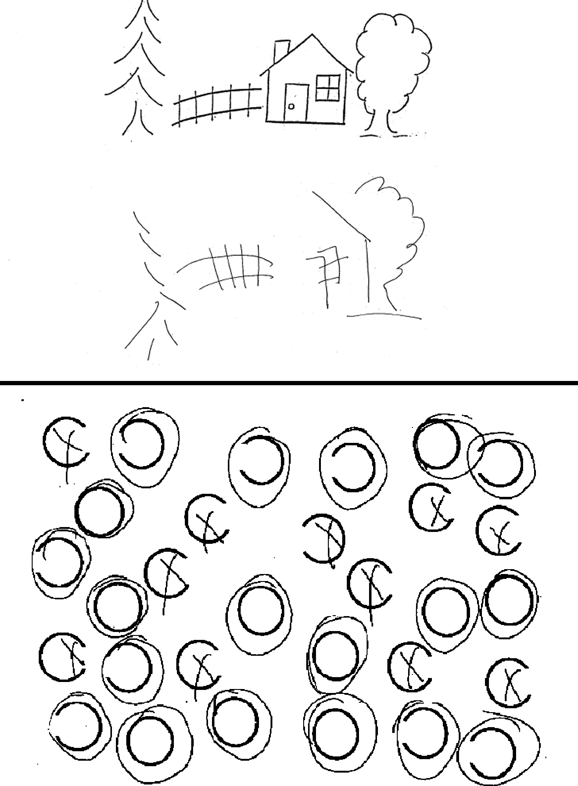
Spatial representations can have reference frames that are relative to the organism (egocentric, e.g., "The coffee cup is left of my body") or relative to objects in space (allocentric, e.g., "The handle is on the left side of the coffee cup"). By studying neurologically intact and impaired individuals, my research has provided evidence regarding the neural correlates of different types of spatial representations. In work with Argye Hillis, we examined the neural correlates of different subtypes of neglect - an attentional deficit subsequent to right hemisphere damage in which individuals fail to respond to stimuli on the left side of space. Previous studies have reported egocentric neglect, in which individuals ignore the contralesional side of space relative to the viewer, and allocentric neglect, in which individuals ignore the contralesional side of each object regardless of its location relative to the viewer. Using perfusion and diffusion weighted imaging (methods used to measure acute brain damage), we found that regions traditionally considered part of the dorsal stream of visual processing were associated with egocentric neglect, whereas ventral stream regions were associated with allocentric neglect (Medina et al., 2009; Khurshid et al., 2011). Furthermore, in collaboration with Roy Hamilton, we are using non-invasive brain stimulation techniques to understand the neural correlates of different types of spatial representations. Transcranial direct current stimulation (tDCS) is a safe, low-cost brain stimulation technique in which a low intensity current runs through the brain. Depending on the electrode configuration, tDCS can focally increase (anodal) or decrease (cathodal) cortical activity. In a recent experiment, we (Medina, Beauvais, et al., 2012) presented neurologically intact individuals with a target identification task, where the target (a circle with a gap) could vary by position in space (left or right side of the array - egocentric) and gap position (left or right side of the circle - allocentric). We found that right anodal tDCS over posterior parietal cortex (a region commonly associated with neglect) resulted in a significant decrease in reaction times for detecting the left side versus the right side of targets (allocentric processing). Guided by our previous work with brain-damage individuals, we are currently using transcranial magnetic stimulation (TMS) to selectively disrupt egocentric versus allocentric processing. Relevant Publications
Our new lab at the University of Delaware has the following resources:
As for future directions, we are using non-invasive brain stimulation techniques, not only to understand relationships between cognitive and neural functioning, but also to see if brain stimulation can enhance cognitive processing both in neurologically intact and impaired individuals. For example, we have recently found that TMS of the intact right hemisphere in individuals with aphasia results in improved discourse productivity on a picture description task (Medina, Hamilton, et al., in preparation). We are also exploring the functional and neural architecture of body representations. Supported by a grant from the University of Delaware Research Foundation, we are presenting brain-damaged individuals with a battery of tests designed to explore dissociations in deficits of body representation, and correlate them with areas of brain damage. Furthermore, we are also using TMS to investigate the neural correlates of subcomponents of the "body schema". In neurologically intact individuals, we are using a novel version of the "mirror box" illusion to understand how the body integrates information from various systems to represent the body. Finally, as new imaging techniques give rise to new statistical analyses for these data, it is important that these analyses be appropriate and meaningful. We have published a paper noting the inappropriate usage of certain statistical tests in voxel-lesion symptom-mapping analyses, which has resulted in large Type I errors in previously published studies (Medina, Kimberg, et al., 2010). We plan on continuing research examining methodological issues in cognitive neuroscience, including best practices for using patient performance to relate structural damage to functional impairments. Relevant Publications
|
